
Knee meniscal tears – how are they often described
 Each meniscus is divided into three sections the anterior horn, the body, and the posterior horn.
The meniscus is then further classified into thirds , being the outer third, middle third and inner third.
When describing meniscal tears, especially based on MRI findings and/or arthroscopic findings, they will usually make reference to the following descriptions:
LOCATION: The tear may be located in the anterior horn, body or posterior horn of the meniscus and may not be exclusive to one area but cross two or three areas. Tears are most common in the posterior horn.
It will then be classified further into which third the tear is located – the outer third, middle third or inner third. Once again the tear may cross over more than one third. This classification is important as will assist in determining the ability of the tear to heal since the blood supply is critical in the healing process, hence tears in the outer third have the best chance of healing.
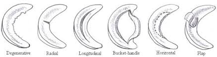
PATTERN: Tears to the meniscus can come in many geometrical patterns such as longitudinal, radial, bucket handle/ displaced bucket handle, parrot beak, flap / displaced flap, horizontal, vertical, degenerative. Complex tear includes more than one pattern.
COMPLETE vs INCOMPLETE: This makes reference to a tear that goes all the way through the meniscus and a piece of tissue is separated from the meniscus is called a complete tear. If the tear is still still partly attached to the body of the meniscus it is called an incomplete tear.
STABLE vs UNSTABLE: A stable tear does not move and may heal on its own, and an unstable tear allows the meniscus to move abnormally and more likely to need surgical intervention.
The classification of meniscal tears helps to determine the best treatment and is important information for the treating practitioner/surgeon as to what path of management to take.