Ice baths – the theoretical benefits

shutterstock_105258182It is a common practice you see, where athletes finish training or an event and immerse themselves in an ice filled bath (cold water immersion) to assist with recovery. Why exactly do they do this and what is the perceived benefit.

Intense exercise leads to microtrauma (small tears) in muscle fibres and resultant soreness which is often referred to as Delayed Onset Muscle Soreness (DOMS). Read more

Stress fractures of the spine in young athletes

cricket-lower-back-pain-300x143Stress fractures of the lumbar spine in the young athlete is a common injury you hear of in this population group. Spondylolysis is a condition describing a non displaced stress fracture of the spinal vertebra, in the pars interarticularis of the spinal column. The pars interarticularis is the part of the vertebra between the upper and lower facets of the vertebra. Most cases of spondylolysis occur at the L5 level.

Read more

Ways to counteract the sedentary nature of sitting in your work day

shutterstock_109552622Prolonged siting in deskbound jobs can definitely have ill effects on our health. Not only does it load the spine, but it also is a very passive activity meaning the level of muscle activity drops and the rate of burning calories is somewhat reduced compared to walking around. Of course though in today’s society many of us have deskbound jobs, so here are some suggested ways to counteract the sitting whilst in the workplace.

Read more

5 easy exercises to help ease tension in your neck

physiotherapy-neck-painFeeling tense in your shoulders and neck? Feel like your shoulders are creeping towards your ears? Do you carry the weight of the world on your shoulders? Try these easy exercises to help alleviate tension in your neck.

  • In a seated or standing position, have your arms by your side, slowly roll your shoulders backwards 10 times.

Read more

Knee meniscal tears – how are they often described

images
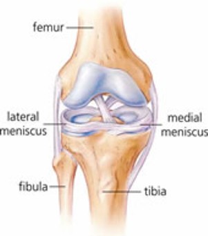
Each meniscus is divided into three sections the anterior horn, the body, and the posterior horn.

The meniscus is then further classified into thirds , being the outer third, middle third and inner third.

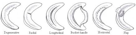
When describing meniscal tears, especially based on MRI findings and/or arthroscopic findings, they will usually make reference to the following descriptions:

LOCATION: The tear may be located in the anterior horn, body or posterior horn of the meniscus and may not be exclusive to one area but cross two or three areas. Tears are most common in the posterior horn.

It will then be classified further into which third the tear is located – the outer third, middle third or inner third. Once again the tear may cross over more than one third. This classification is important as will assist in determining the ability of the tear to heal since the blood supply is critical in the healing process, hence tears in the outer third have the best chance of healing.

Read more

What is Knee meniscus?

knee-meniscusThe knee joint is made up of three bones, the femur (thigh bone), the tibia (shin bone) and the patella (knee cap). The knee joint is a hinge joint allowing two principal actions flexion (bending) and extension (straightening). The meniscus is “C shaped” discs made of tough cartilage called fibrocartilage. They are located on the tibial plateau (top surface of the tibia) between the tibia (shin bone) and the femur (thigh bone).

There is a lateral meniscus which is located on the outside of the knee joint, and a medial meniscus located on the inside of the knee joint. The meniscus plays a role in joint stability by creating a cup for the femur to sit in, and also act as a shock absorber for the knee by spreading compression forces from the femur over a wider area on the tibia. This function helps to protect the underlying joint surface cartilage that lines the bone surfaces from wearing out and developing degenerative arthritis.

Read more